- About Us
- Meet The Experts
- Patient Information
- Referring Doctors
- Seminars & Lectures
- Reviews
- Contact Us
Need Immediate Assistance?
Speak directly with our team.
Speak directly with our team.
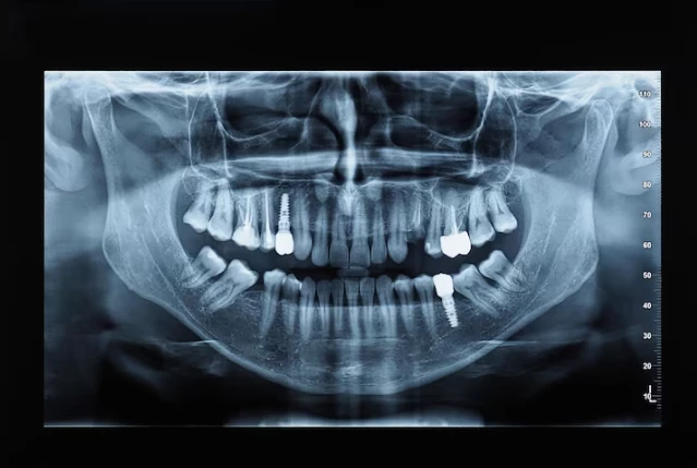
Gain a complete view of your oral health with advanced 3D Cone Beam CT (CBCT) imaging. Our in-office technology provides highly detailed images of your teeth, jawbone, nerves, sinuses, and surrounding structures to support accurate diagnosis and precise treatment planning.
Successful oral surgery begins with accurate diagnosis and treatment planning. CBCT technology provides detailed information that cannot always be seen on standard dental X-rays.
Benefits of CBCT Imaging:
By providing a comprehensive view of your oral anatomy, CBCT imaging helps your surgeon make informed treatment decisions and develop a personalized surgical plan designed around your specific needs.
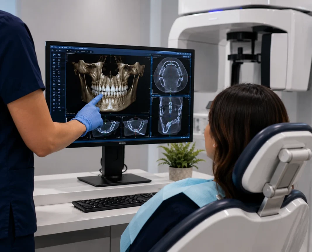
Our oral surgeons may recommend Cone Beam CT imaging for a variety of diagnostic and treatment planning needs. The detailed 3D images help support more accurate evaluations and personalized care.
Evaluate bone volume, density, and implant positioning for long-term success.
Assess tooth position and proximity to nerves before extraction.
Identify critical structures to support safer surgical procedures.
Measure available bone and evaluate sinus anatomy before treatment.
Detect bone loss, structural concerns, and areas requiring reconstruction.
Assist in evaluating cysts, lesions, infections, and other oral health concerns.
Provide detailed imaging for advanced surgical planning and treatment.
Visualize jaw joint anatomy and identify structural conditions that may affect function and treatment planning.
Evaluate airway structures and surrounding anatomy to support comprehensive diagnosis and treatment planning.
CBCT scans are performed right here in our office, eliminating the need for outside imaging centers.
The detailed information obtained from CBCT imaging allows us to provide more personalized and precise surgical care, helping ensure optimal outcomes for every patient.
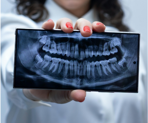
Getting a Cone Beam CT scan is quick, comfortable, and completely non-invasive. The entire process typically takes only a few minutes.
You will stand or sit comfortably while the scanner is carefully positioned around your head.
The scanner rotates around your head for a few seconds while capturing highly detailed images.
Advanced software processes the scan and creates a detailed three-dimensional model of your anatomy.
Your surgeon reviews the scan and develops a personalized treatment plan based on your unique anatomy and needs.
Discover how advanced 3D Cone Beam CT imaging helps us provide safer, more accurate, and more personalized oral surgery treatment.
Call Our Office NowOur advanced Cone Beam CT technology provides detailed 3D imaging that helps ensure accurate diagnosis, precise treatment planning, and exceptional patient care.
State-of-the-art 3D imaging equipment provides highly detailed views for accurate diagnosis and treatment planning.
Detailed evaluation before dental implants, extractions, bone grafting, and other oral surgery procedures.
Treatment recommendations are tailored to your unique anatomy, oral health needs, and long-term goals.
Advanced imaging is interpreted by experienced oral and maxillofacial surgical professionals.
CBCT scans are completed right in our office, eliminating the need for additional appointments elsewhere.
Immediate access to detailed scan data allows for faster consultations and more streamlined care.
See what patients have to say about their experience with Glen Burnie Oral Surgery.
Based on hundreds of positive patient experiences and trusted referrals throughout the region.
Learn more about Cone Beam CT imaging, how it works, and why it plays an important role in oral surgery diagnosis and treatment planning.
At Glen Burnie Oral Surgery Associates we proudly provide advanced oral surgical procedures, wisdom teeth removal, dental implants , all - on-4 implants, IV sedation and emergency oral surgery.